Anterior View of the Pelvis and Hip
Services
Welcome to the detailed exploration of the anterior view of the pelvis and hip by Shout It Marketing, your trusted source for Business and Consumer Services - Digital Marketing. In this comprehensive guide, we will delve into the intricacies of the pelvis and hip anatomy from an anterior perspective.
Understanding the Pelvis and Hip
The anterior view of the pelvis provides a unique perspective on the structure and function of this vital part of the human body. The pelvis is a bony structure located at the base of the spine, serving as the foundation for the hip joints and supporting the weight of the upper body. It consists of several bones, including the ilium, ischium, and pubis, which fuse together to form the pelvic girdle.
Anatomy of the Pelvis
When examining the anterior view of the pelvis, key structures come into focus, such as the acetabulum, the socket of the hip joint, and the symphysis pubis, a cartilaginous joint connecting the pubic bones. The pelvis also houses the pelvic organs, including the bladder, reproductive organs, and part of the digestive system.
Bones of the Pelvis
The ilium, ischium, and pubis are the three primary bones that make up the pelvis. The ilium represents the uppermost and largest bone, while the ischium forms the lower and posterior part. The pubis completes the pelvic circle in the front.
Joints of the Pelvis and Hip
The pelvis is connected to the spine through the sacroiliac joint, allowing for stability and mobility. The hip joint, also known as the acetabulofemoral joint, is a ball-and-socket joint that enables a wide range of motion in the lower limbs.
Importance of the Anterior View
Studying the anterior view of the pelvis is crucial for various medical professionals, including orthopedic surgeons, physical therapists, and radiologists. It provides valuable insights into diagnosing and treating conditions affecting the pelvis and hip region.
Common Pathologies and Conditions
From fractures and dislocations to osteoarthritis and avascular necrosis, several conditions can impact the pelvis and hip. Understanding the anatomy from an anterior perspective aids in identifying and managing these issues effectively.
Diagnostic Techniques
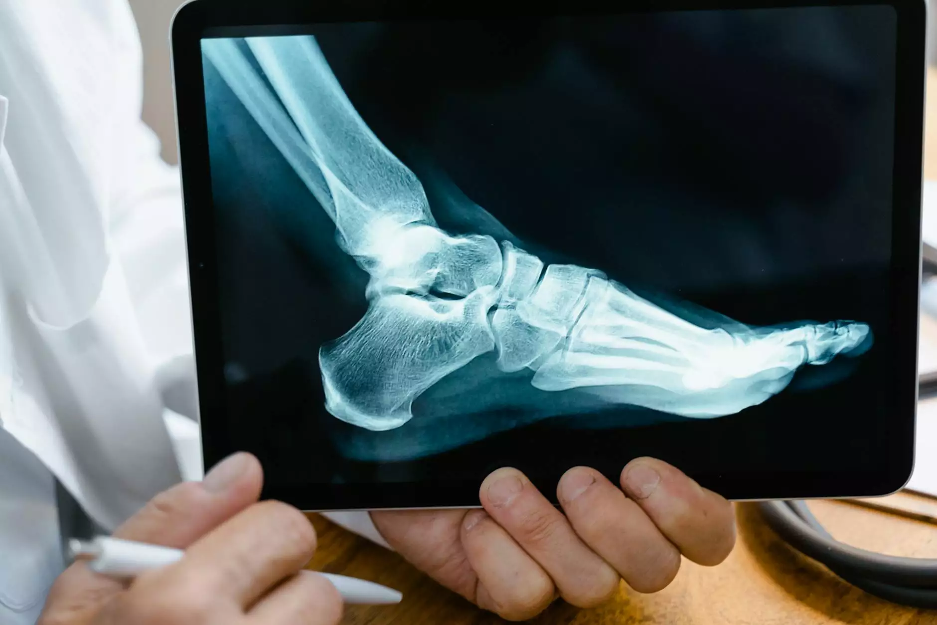
Medical imaging modalities like X-rays, CT scans, and MRIs play a crucial role in visualizing the anterior view of the pelvis and hip. These diagnostic tools help healthcare professionals assess the structural integrity and function of these interconnected regions.
Conclusion
In conclusion, exploring the anterior view of the pelvis and hip provides a deeper understanding of the complex anatomical relationships within this region. Whether you are a healthcare professional or a curious individual seeking knowledge, this guide by Shout It Marketing aims to enrich your insights into the fascinating world of human anatomy.


