Bony Landmarks of the Knee: Posterior View
Services
Welcome to Shout It Marketing's detailed guide on the bony landmarks of the knee from a posterior view. Understanding the anatomy and structure of the knee is crucial for various fields such as medicine, physical therapy, and sports science. In this comprehensive overview, we will delve into the key bony landmarks that make up the knee joint.
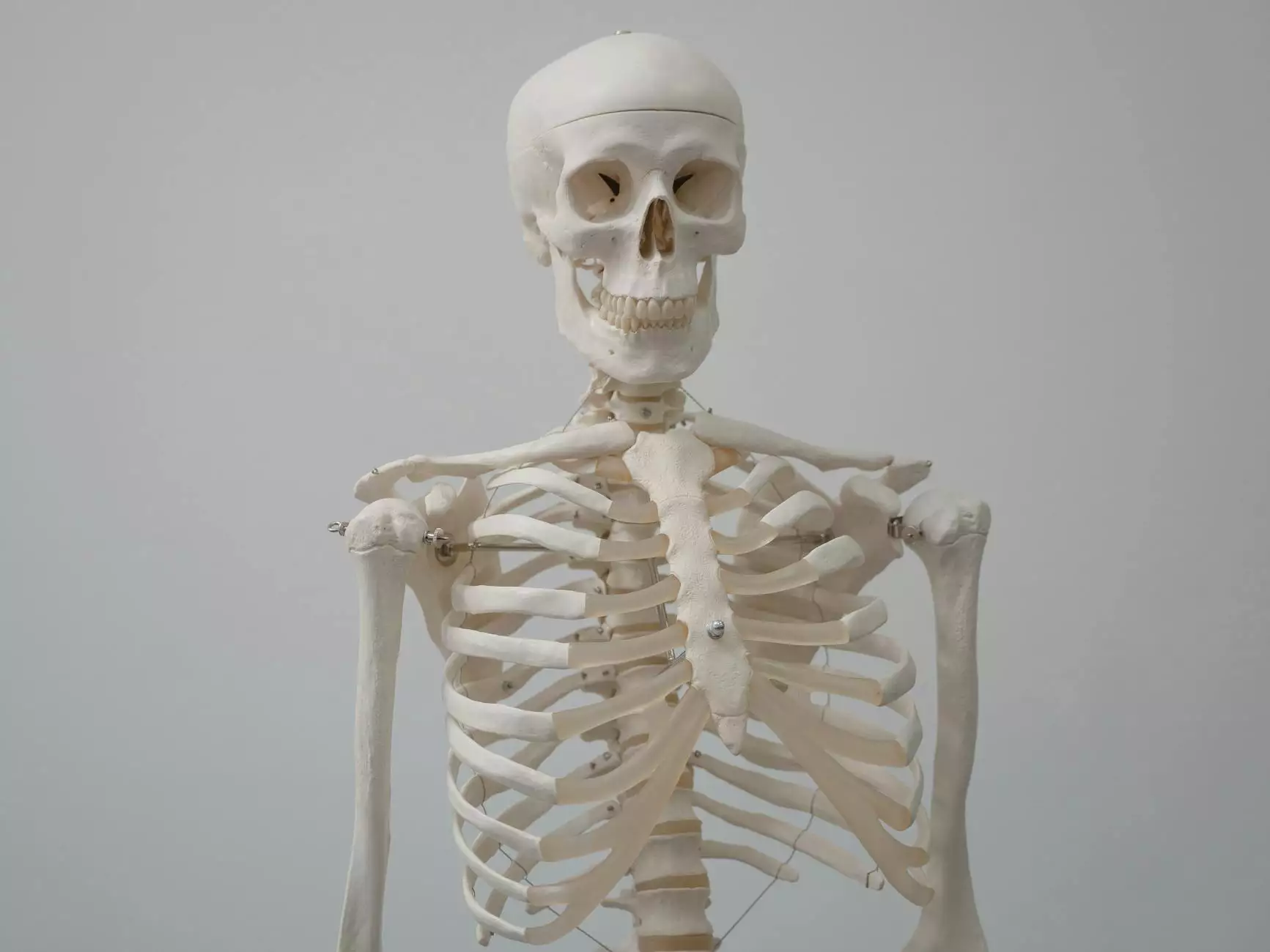
Anatomy of the Knee
The knee joint is one of the largest and most complex joints in the human body. It is formed by the articulation of three bones: the femur (thighbone), the tibia (shinbone), and the patella (kneecap). The knee joint is supported by a network of ligaments, tendons, and muscles that work together to provide stability and mobility.
Bony Landmarks
When viewing the knee from the posterior aspect, several important bony landmarks can be identified:
Femur
- The femoral condyles are the rounded projections at the distal end of the femur that articulate with the tibia to form the knee joint.
- The intercondylar fossa is a groove located between the femoral condyles that accommodates the cruciate ligaments of the knee.
Tibia
- The tibial plateau is the top surface of the tibia that articulates with the femoral condyles.
- The tibial tuberosity is a bony prominence on the anterior aspect of the tibia where the patellar ligament attaches.
Patella
- The patellar surface is the smooth, anterior surface of the patella that glides over the femoral condyles during knee movement.
- The apex of the patella is the pointed inferior portion of the bone.
Function of Bony Landmarks
Each bony landmark of the knee plays a crucial role in the function and stability of the joint. The femoral condyles and tibial plateau provide the articulation points for flexion and extension movements, while the ligaments and tendons attached to the tibial tuberosity and patella help to stabilize the joint during weight-bearing activities.
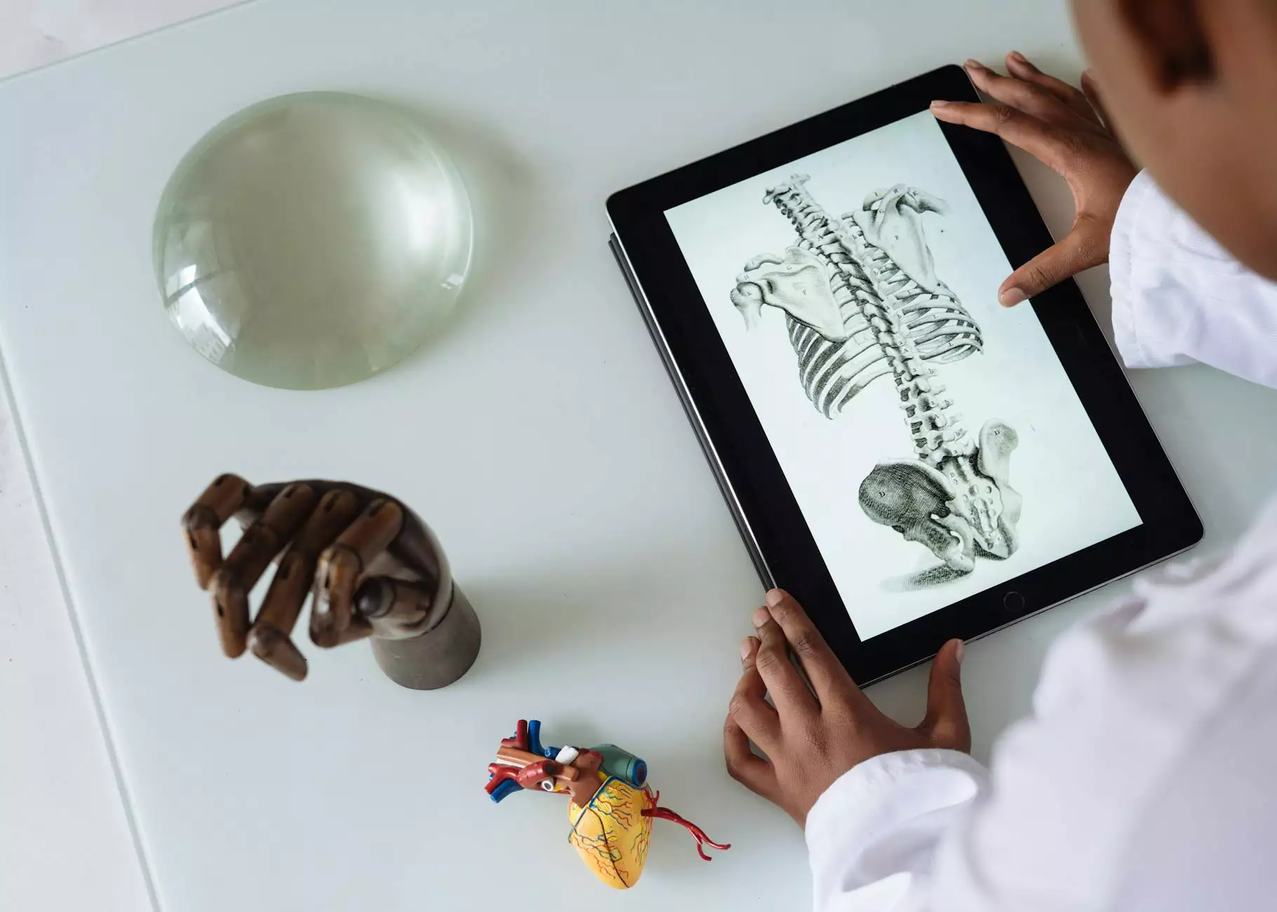
Clinical Significance
Understanding the bony landmarks of the knee from a posterior view is essential for diagnosing and treating various knee injuries and conditions. Healthcare professionals use this knowledge to identify specific areas of the knee joint during physical examinations, imaging studies, and surgical procedures.
Conclusion
In conclusion, the bony landmarks of the knee from a posterior view are integral to the overall structure and function of the knee joint. By gaining a thorough understanding of these anatomical features, individuals in the fields of medicine, physical therapy, and sports science can enhance their knowledge and skills in assessing and managing knee-related issues.